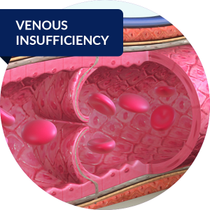
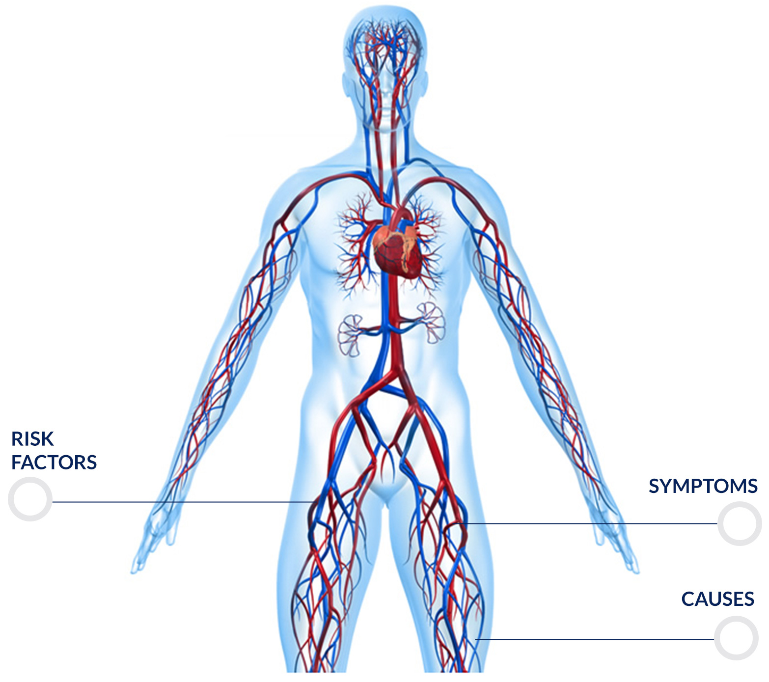
 Venous disease is a clinical condition characterized by a difficult return of blood from the lower limbs to the heart, which occurs when the venous wall and/or valves in the veins of the legs do not function effectively. It affects up to 80% of the population, with a female/male ratio of 3/1.
Select to learn more

Symptoms
The severity of CVI and the complexity of treatment can facilitate disease progression. This is why it is essential that you speak to your doctor if you suffer from any of the symptoms of CVI. The issue will not go away on its own; the sooner it is diagnosed and treated, the higher the chance to avoid severe complications.
Symptoms include:
- Swelling of the legs and ankles, especially after standing up for a long time
- Tired legs
- New varicose veins
- Skin that looks like leather
- Flaking and itching of the skin on the legs or feet
- Stasis ulcers (or venous stasis ulcers)
If left untreated, CVI can swell up and increase venous pressure in the veins, until smaller blood vessels in the legs (capillaries) start bursting. This could turn your skin reddish-brown, and make it more sensitive to breakage caused by trauma or scratches. It can cause inflammation of the nearby tissue and damage internal tissues. In more severe cases, this can lead to ulcers, open wounds on the surface of the skin. These venous stasis ulcers can be hard to treat and can become infected. When the infection is not kept under control, it can spread to the surrounding tissue causing a condition known as cellulitis.
CVI is often associated with varicose veins, which causes tangled or enlarged vein near the surface of the skin. This can occur anywhere, but most commonly affects the legs.
Causes
Veins are blood vessels carrying the blood back to the heart from all organs in the body. In order to reach the heart, blood must flow upwards through the veins in the legs. Muscles in the calf and foot must contract at each step in order to compress veins to push the blood upwards; thanks to this and two other mechanisms devoted to the same function, vis a tergo (strength imparted by arteries to push blood) and the heart’s churning effect, blood reaches the heart from the periphery of the body, particularly – in this case – from the lower limbs. Veins contain one-way valves that keep blood flowing against gravity.
Chronic venous insufficiency occurs when these valves are damaged, allowing the blood to flow backward. Valves can suffer damage due to aging, a sedentary lifestyle or one that involves standing for long periods of time (e.g. specific types of jobs) or a combination of aging and reduced mobility. When veins and valves are weakened to the point where it becomes difficult for blood to flow up all the way to the heart, venous blood pressure remains high for long periods of time, causing chronic venous insufficiency.
CVI is usually a result of a blood clot forming in the deep veins in the leg (a condition known as deep vein thrombosis (DVT)). CVI can also be caused by pelvic tumours or vascular malformations, but sometimes its causes are unknown. Valve insufficiency in the veins of the legs causes the blood flow in the veins to slow down, causing the legs to swell.
Chronic venous insufficiency that develops as a consequence of DVT is also known as post-thrombotic syndrome. As many as 30% of people suffering with DVT will develop this condition in the 10 years following diagnosis.
Risk factors
If you are at risk of CVI, you may be more likely to develop the disease. The main risk factors are:
- Deep vein thrombosis (DVT)
- Varicose veins or a family history of varicose veins
- Obesity
- Pregnancy
- Lack of exercise
- Smoking
- Standing for long periods of time or a sedentary lifestyle
- Being a woman
- Being over 50 years of age
Around 40% of people in the United States are estimated to suffer with CVI. It occurs more frequently in people over the age of 50 and it is more common in women.
How is CVI diagnosed?
Deep vessel doppler ultrasonography
In order to make a diagnosis of CVI, your doctor will look at your medical history and perform a physical examination. During this, the doctor will carefully analyse your legs.
A test called vascular or doppler ultrasonography can be used to examine the blood flow in the legs. During a vascular ultrasound, a transductor (a small portable device) is placed on the skin above the vein that will be examined. The transductor releases sound waves which bounce back from the vein. These sound waves are registered and an image of the flow is shown on a screen.


